Star

Home

Biochem

Biochem

CellBio

CellBio

Genetics

Genetics

Orf

Orf

Hydro

Hydro

Molsim

Molsim

Cluster

Cluster

Hpc

Hpc

Contents (Version 2)


  1. Welcome to StarBiochem
  2. Opening StarBiochem
  3. StarBiochem at a glance
  4. Loading a protein structure
  5. Manipulating the default view
  6. Visualizing atoms and bonds
  7. Displaying molecular structures
  8. Additonal Features

Welcome to StarBiochem

StarBiochem is a 3D protein viewer. StarBiochem runs on standard Linux, Windows and Mac computers.

Return to top

Opening StarBiochem

To get started with StarBiochem:

  1. Navigate to http://web.mit.edu/star/biochem.

  2. Click on the Start button to launch the StarBiochem application.

  3. Click Trust when a prompt appears asking if you trust the certificate.

Return to top

StarBiochem at a glance

This is the view you will see when you open StarBiochem.
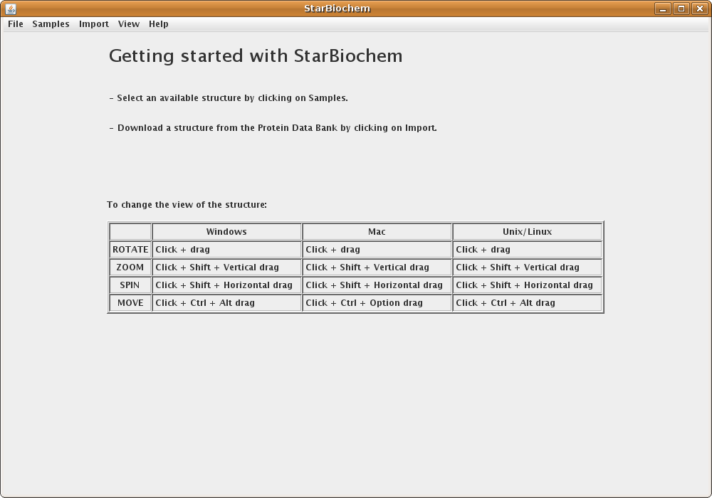
StarBiochem Getting Started Screenshot

Return to top

Loading a protein structure

StarBiochem enables the visualization of molecules encoded within the Protein Data Bank (PDB) files. PDB files are named by a four-character ID such as “1GXX”. You may load files directly from the PDB, the Samples menu or open files stored locally on your computer.

  1. Click on Import -> RCSB (Protein Data Bank).

  2. Enter the four-character ID of a PBD file (eg. 3D9S), click Open.
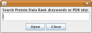
    Search Protein Data Bank

  3. When the search results appear, choose the desired PDB file and click Open.
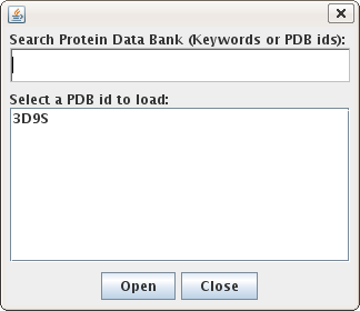
    Select Protein Data Bank from Search

  • Alternatively, select a PDB file from list found in the Samples menu.

  • To load a PDB file stored locally on your computer, click on File -> Open and select the desired file.

Upon opening a PDB file in StarBiochem, you will see a 3D model of the protein in the viewer.
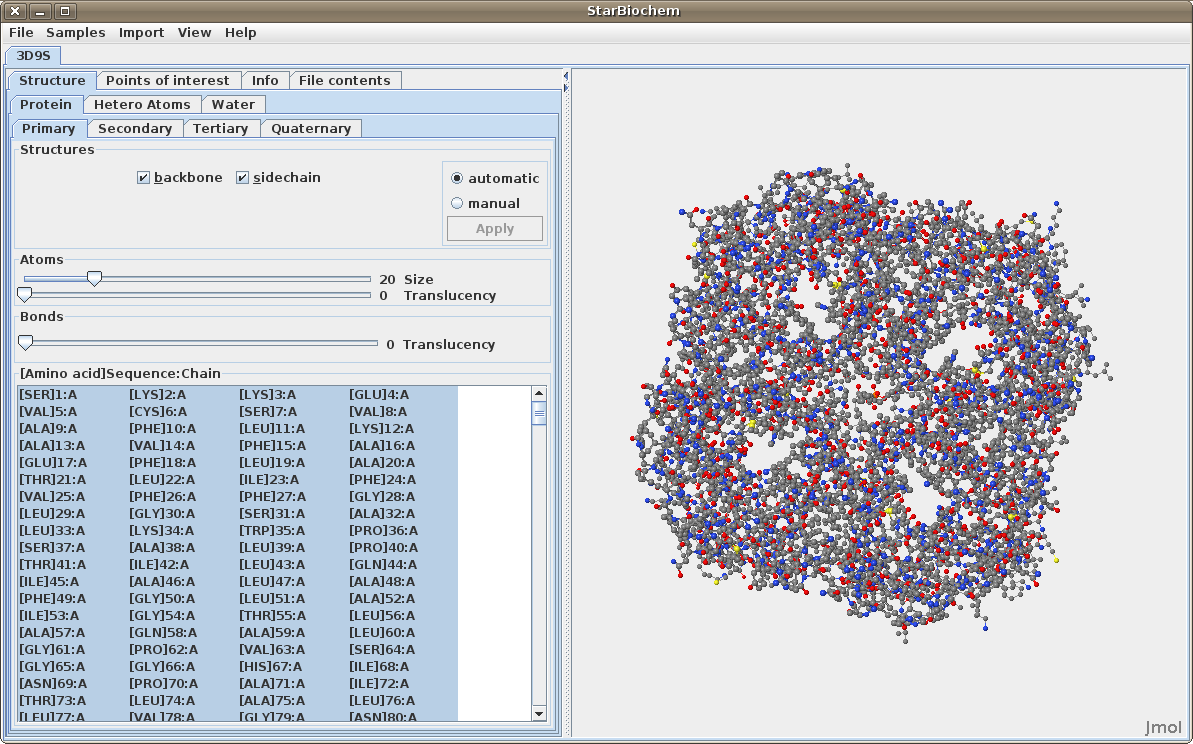
StarBiochem Default View

Note:
Each tab contains Automatic and Manual options which allow you to choose when the changes made are applied to the image.

  • Automatic immediately applies selection, size or translucency changes.
  • Manual allows you to make mutiple changes before clicking the Apply button. This is usually helpful when isolating a specific portion of a structure or making mutiple changes to a large structure which may require more time to render.

When multiple PBD files are open, they can be closed individually or all at once by clicking on File -> Close, or File -> Close All respectively.

Return to top

Manipulating the default view.

To change the view of the structure:

Change the view of structure

Return to top

Visualizing atoms and bonds

There are two different 3D models that are used by most protein 3D viewers, including StarBiochem, to visually represent the structure of molecules:

ball-and-stick model: the atom's nucleus is drawn as a ball and covalent bonds connection atoms are drawn as lines.

  • Advantage: allows a better view of how atoms bond together.

space-filled model: atoms are drawn as spheres, whose size represent the physical space an atom occupies (the nucleus plus its surrounding electrons, also called van der Waals radius).

  • Advantage: allows for a more accurat representation of the size of atoms and molecules.

You can switch from the ball-and-stick model to the space-filled model in StarBiochem by increasing the size render of the atoms in the structure.
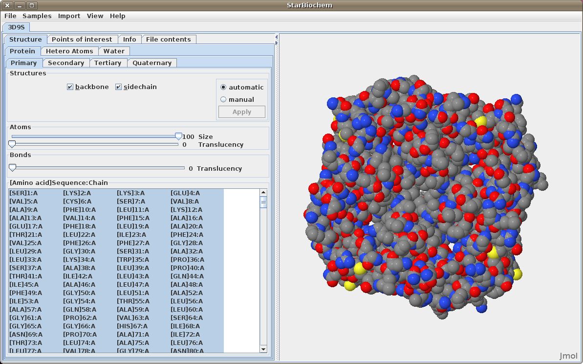
  • Click on the Primary tab. By default the atom size is 20% which represents the ball-and-stick model. Move the Atoms size slider completely to the right, 100%, to switch to the space-filled model.

Space Fill View StarBiochem

The default coloring scheme for atoms is as follows:

  • Carbon (C) atoms grey spheres
  • Oxygen (O) red spheres
  • Nitrogen (N) blue spheres
  • Sulfur (S) yellow spheres

(Hydrogen atoms are shown as white though they are rarely depicted)

Return to top

Displaying molecular structure

The primary structure of a protein lists the amino acids that make up a protein’s sequence or the nucleotides that make up a DNA segment, but does not describe their corresponding shape. The secondary structure of a protein refers to how stretches of amino acids within a protein chain are arranged in space in characteristic patterns such as helices, sheets, and coils. The tertiary structure describes the folded shape of a protein chain and is determined by characteristic properties of the amino acid residues, such as acidic, basic, polar, and non-polar. The quaternary structure indicates the relationship between the chains of a molecule.

Displaying primary structures:
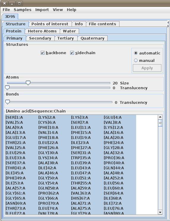
  • Click on the Primary tab.

Primary Structures

  • This view shows a numbered list of amino acids and options for viewing the backbone and sidechain. By default all of the amino acids are selected.
  • Amino acids consist of two parts, backbone and sidechain. Unchecking the box beside backbone or sidechain allows changes to apply only the part that is checked.

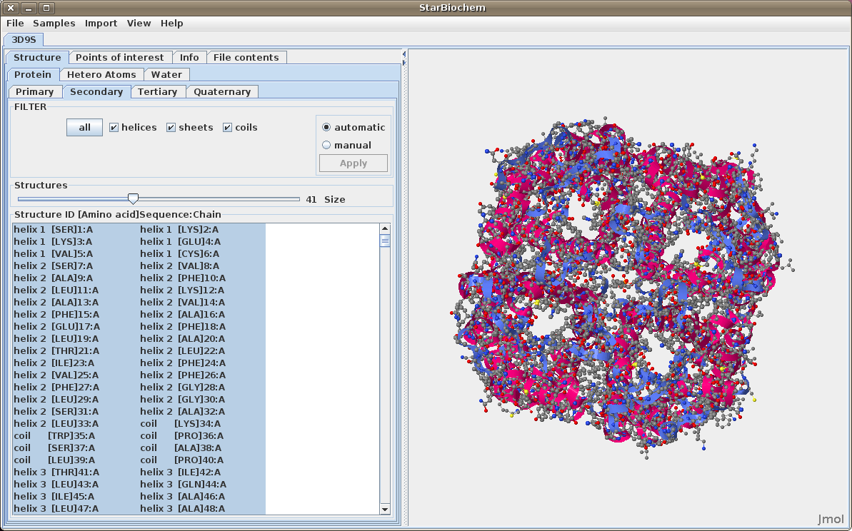
Displaying secondary structures:

  • Click on the Secondary tab.

Secondary Structures

  • This view shows a numbered list of the helices, sheets and/or coils with their amino acids. By default the structures size is set to zero.
  • To display the secondary structures move the Size slider to the right to increase the size of the structure(s). Check or uncheck the box beside each structure as needed.

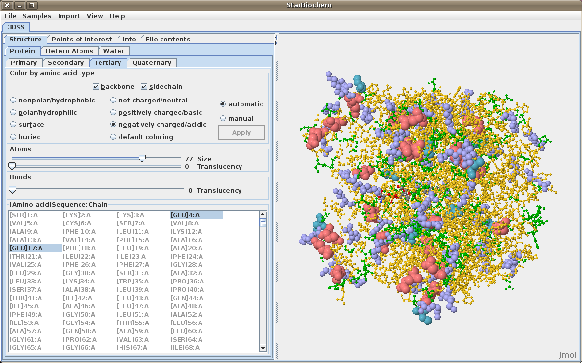
Displaying tertiary structures:

  • Click on the Tertiary tab.

Tertiary Structures

  • This view shows a numbered list of amino acids. The default view will mirror the selections and sizes from the Primary tab.
  • To display the residue colors click on the buttons beside the amino types, e.g. polar, nonpolar, etc.
  • Click on the default coloring button to return to the default coloring scheme.

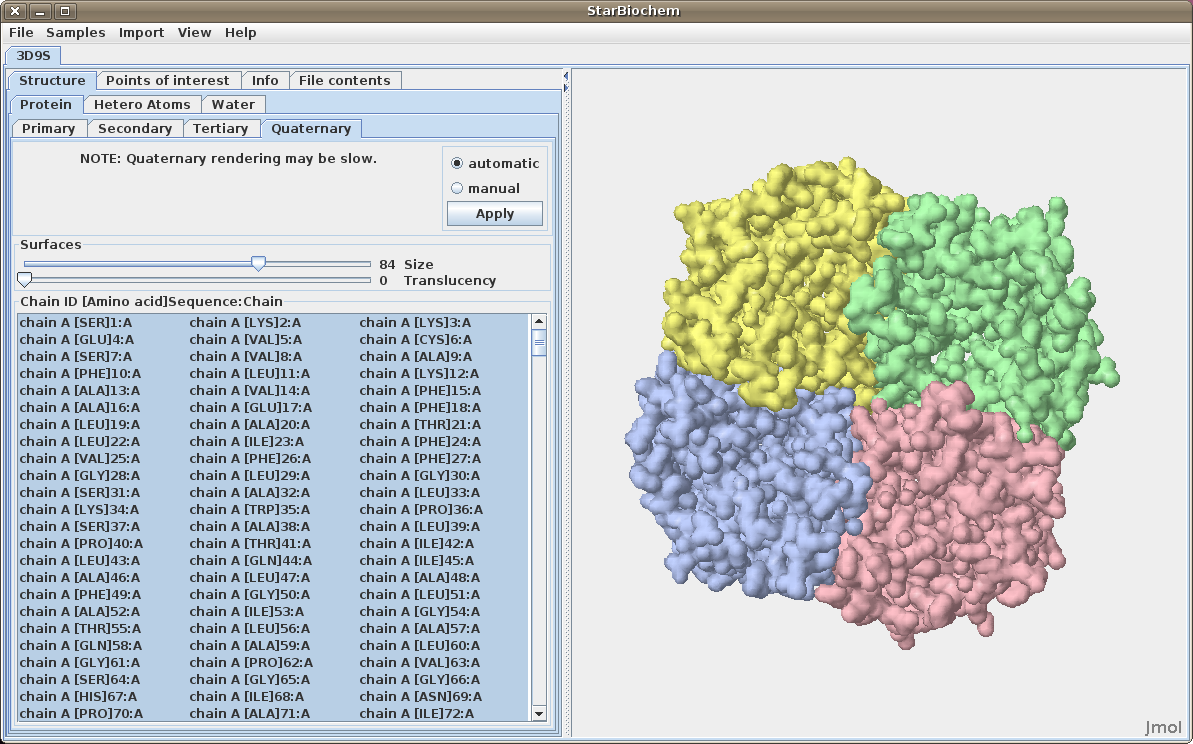
Displaying quaternary structures:

  • Click on the Quaternary tab.

Quaternary Structures

  • This view will list the quarternary chains which are alphabetically or numerically identified (e.g. chain A, chain B). By default the surfaces' size is set to zero.
  • To display the quarternary structures (chains) increase the size by moving the size slider to right.

Displaying nucleic acids:

Click on the Nucleic Acids tab.
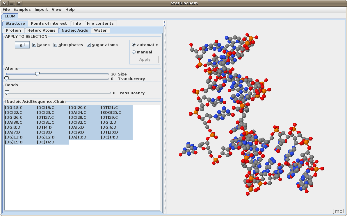
Nucleic Acids

  • This view will show a list of the nucleic acids and options for selecting some or all of the nucliec structures (bases, phosphates, or sugars).

Return to top

Selecting Atoms and Structures

StarBiochem allows for selection of individual or groups of atoms and structures.

Selecting Sequences (Primary, Secondary and Tertiary):

  • To select a single item in the sequence list simply click on it.
  • To select multiple items individually, click on the first item, press Ctrl (PC/Linux) or Apple (Mac) and click on the remaining sequences desired.
  • To select multiple amino acids as a group click on the first amino acid, press Shift and click on the last amno acid.
  • To select multiple groups of amino acids, click on the first amino acid, press Shift and click on the last amino acid in the first group. Then press Ctrl (PC/Linux) or Apple (Mac) and click on the first amino acid of the next group, press Ctrl (PC/Linux) or Apple (Mac)+Shift and click on the last amino acid of the group.

Selecting Quaternary Sequences:

  • Click on a list entry to select a particular chain in the viewer. The Quaternary tab does not allow partial chain selections.
  • To select multiple chains, press Ctrl while clicking on each chain.
  • Adjust the size slider under Surfaces to change the amount of surface shown.
  • Move the translucency slider to the right under Surfaces to reveal the amino acids beneath the surfaces.

Additional Features

Displaying water atoms

Some proteins have the addition of a Water tab. Select this tab to see the oxygens that make up water molecules within the protein (hydrogens rarely defined).
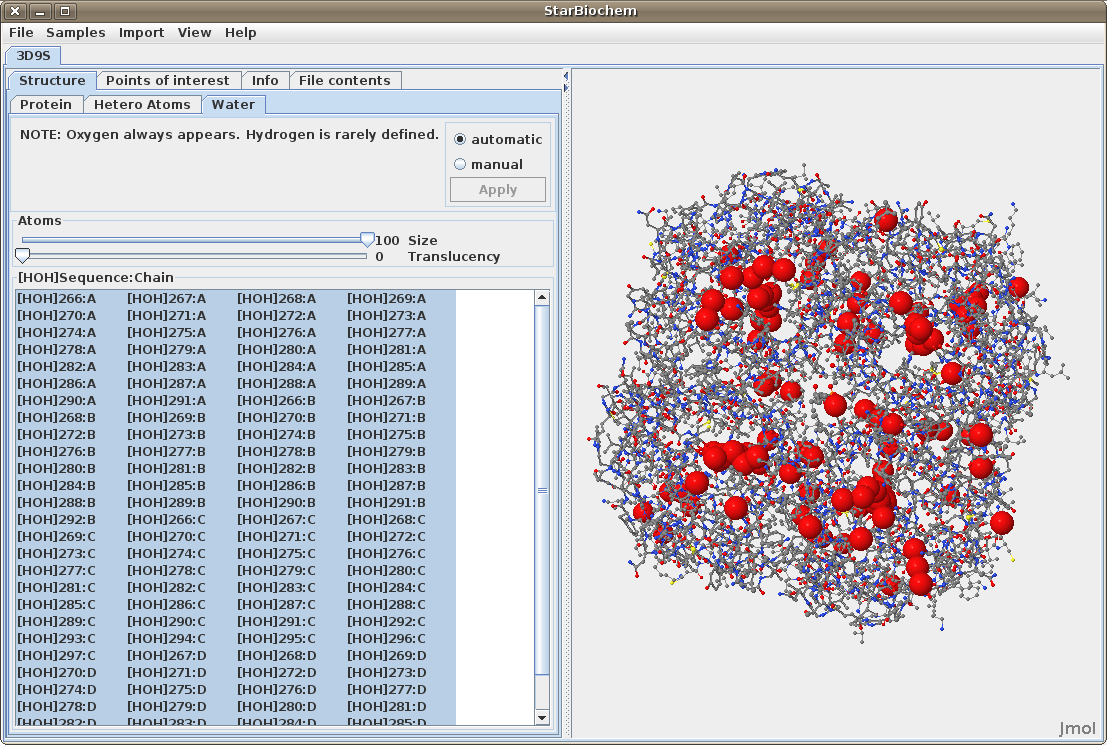
Water

Displaying hetero atoms
Some proteins have the addition of a Hetero Atoms tab. Select this to view any hetero atoms within the protein and change their size in the viewer.
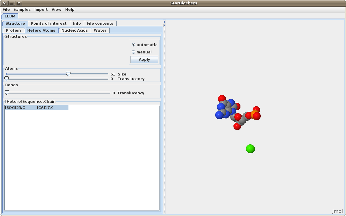
Hetero Atoms


Return to top